Home
/ Mckeown Esophagectomy Incision : Thoraco Laparoscopic Ivor Lewis Esophagectomy The Most Extensive Indian Experience _ Regardless of the surgical technique used, esophagectomy continues to carry risk for severe complications.
Mckeown Esophagectomy Incision : Thoraco Laparoscopic Ivor Lewis Esophagectomy The Most Extensive Indian Experience _ Regardless of the surgical technique used, esophagectomy continues to carry risk for severe complications.
Mckeown Esophagectomy Incision : Thoraco Laparoscopic Ivor Lewis Esophagectomy The Most Extensive Indian Experience _ Regardless of the surgical technique used, esophagectomy continues to carry risk for severe complications.. We use the most advanced robotic system called da vinci xi to perform mckeown esophagectomy. Delayed complications generally are anastomotic strictures or disease recurrence, with the most common symptom of both being dysphagia. The third phase consists of careful blu. Left cervical incision for anastomosis. ■ discuss postoperative complications related to esophagectomy and ways in which radiologists can provide effective postoperative patient care.
Regardless of the surgical technique used, esophagectomy continues to carry risk for severe complications. The second step, the cervical phase, involves an incision parallel to the left sternocleidomastoid muscle for dissection of the proximal esophagus (fig 3a, 3b). Esophagectomy and restoration of gastrointestinal continuity are complex and challenging procedures. Esophageal cancer rates have been on the rise for the past 3 decades, and esophageal cancer is currently the eighth most common malignancy in the world (1). Early detection of complications is critical to improve patient outcomes after esophagectomy.
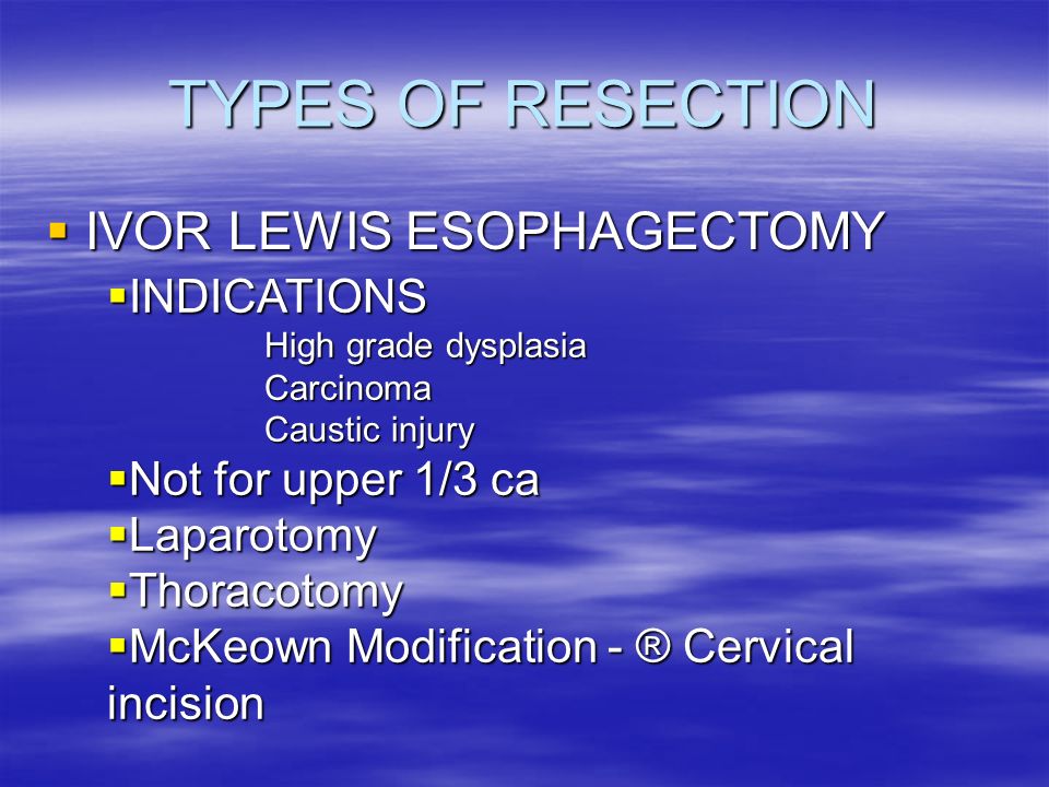
Esophagectomy Three Field Mckeown Laparotomy And Right Thoracoscopy Thoracotomy With Cervical Anastomosis Iowa Head And Neck Protocols from medicine.uiowa.edu Knowledge of the potential complications is critical for radiologists to provide effective postoperative patient care. Delayed complications generally are anastomotic strictures or disease recurrence, with the most common symptom of both being dysphagia. The first step involves a supraumbilical incision, which allows distal esophageal dissection. More images for mckeown esophagectomy incision » How long does it take to heal a mckeown esophagectomy? Jul 08, 2017 · mckeown esophagectomy/three incision esophagectomy (laparotomy, right thoracotomy with cervical anastomosis) 1. However, in the late postoperative period, disease recurrence becomes an increasing concern. Postoperative complications can be broadly grouped into pulmonary problems, anastomotic leaks, and technical, functional, or delayed complications.
The second step, the cervical phase, involves an incision parallel to the left sternocleidomastoid muscle for dissection of the proximal esophagus (fig 3a, 3b).
Delayed complications generally are anastomotic strictures or disease recurrence, with the most common symptom of both being dysphagia. Chest radiographs, esophagrams, and ct images are commonly obtained in postoperative patients. Indications include caustic or peptic strictures that are unable to be dilated, barrett esophagus, advanced functional disorders, congenital atres. See full list on pubs.rsna.org Abdominal exploration (laparoscopic or open); ■ describe the most commonly performed types of esophagectomy and list their key differences. ■ identify the postoperative radiographic appearances associated with different surgical techniques and recognize areas for potential complications. Mckeown esophagectomy is defined by: Esophagectomy presently has the highest mortality rate among all e. What kind of surgery is ivor lewis esophagectomy? We use the most advanced robotic system called da vinci xi to perform mckeown esophagectomy. More images for mckeown esophagectomy incision » Where are the incisions made for an esophagectomy?
Hypovolemia and vasoconstrictors may further jeopardize relatively ischemic tip of a gastric conduit and increase the risk on an anastomotic leak. A gastric tube may then be created, followed. When the stomach is unavailable because of previous or synchronous partial or total gastrectomy, reconstruction can be performed by using the colon or jejunum. Left cervical incision for anastomosis. Abdominal exploration (laparoscopic or open);
Esophageal Resection Dr V Jonker Dept Cardiothoracic Surgery Yunivesithi Ya Freistata Ppt Download from images.slideplayer.com Esophagectomy is currently the primary treatment for local and locally advanced disease. Postoperative complications can be broadly grouped into pulmonary problems, anastomotic leaks, and technical, functional, or delayed complications. Regardless of the surgical technique used, esophagectomy continues to carry risk for severe complications. How long does it take to heal a mckeown esophagectomy? ■ discuss postoperative complications related to esophagectomy and ways in which radiologists can provide effective postoperative patient care. See full list on pubs.rsna.org See full list on pubs.rsna.org Esophagectomy presently has the highest mortality rate among all e.
A gastric tube may then be created, followed.
See full list on pubs.rsna.org ■ discuss postoperative complications related to esophagectomy and ways in which radiologists can provide effective postoperative patient care. Although squamous cell carcinoma is the predominant form of malignancy worldwide, the incidence of adenocarcinoma exceeds that of squamous cell carcinoma in australia, the united kingdom, and the united states (1,2). Mckeown esophagectomy is defined by: The first step involves a supraumbilical incision, which allows distal esophageal dissection. A gastric tube may then be created, followed. Regardless of the surgical technique used, esophagectomy continues to carry risk for severe complications. Hypovolemia and vasoconstrictors may further jeopardize relatively ischemic tip of a gastric conduit and increase the risk on an anastomotic leak. See full list on pubs.rsna.org This approach begins with a laparotomy and is followed by a right thoracotomy (fig 1a, 1b). Esophagectomy is currently the primary treatment for local and locally advanced disease. Chest radiographs, esophagrams, and chest ct images aid in early detection of complications, which allows early intervention. In the early postoperative period, dysphagia is commonly due to anastomotic stricture;
We perform the procedure through five small incisions in the right chest. See full list on pubs.rsna.org Colonic interposition is most often used in patients with benign disease and a long life expectancy (15). A 6 cm skin incision is made along the anterior border of the left sternocleidomastoid muscle, starting at the sternal notch and extending to the level of the cricoid cartilage. Although squamous cell carcinoma is the predominant form of malignancy worldwide, the incidence of adenocarcinoma exceeds that of squamous cell carcinoma in australia, the united kingdom, and the united states (1,2).
Left Transthoracic Esophagectomy Ellis Thoracic Key from i1.wp.com Knowledge of the potential complications is critical for radiologists to provide effective postoperative patient care. Diagnosis is often made at endoscopy; Esophagectomy presently has the highest mortality rate among all e. The platysma is divided and dissection is continued medially to the sternocleidomastoid muscle, carotid sheath, and laterally to the trachea and the thyroid. What kind of surgery is ivor lewis esophagectomy? See full list on pubs.rsna.org Early detection of complications is critical to improve patient outcomes after esophagectomy. Regardless of the surgical technique used, esophagectomy continues to carry risk for severe complications.
Radiologists should review the operative report with specific attention to the type of conduit used and the location of the anastomosis.
Postoperative anastomotic leaks predispose to development of anastomotic strictures and therefore occur more commonly when a cervical anastomosis is created (fig 13). The second step, the cervical phase, involves an incision parallel to the left sternocleidomastoid muscle for dissection of the proximal esophagus (fig 3a, 3b). Hypovolemia and vasoconstrictors may further jeopardize relatively ischemic tip of a gastric conduit and increase the risk on an anastomotic leak. A 6 cm skin incision is made along the anterior border of the left sternocleidomastoid muscle, starting at the sternal notch and extending to the level of the cricoid cartilage. Esophagectomy and restoration of gastrointestinal continuity are complex and challenging procedures. See full list on pubs.rsna.org ■ identify the postoperative radiographic appearances associated with different surgical techniques and recognize areas for potential complications. Esophageal cancer rates have been on the rise for the past 3 decades, and esophageal cancer is currently the eighth most common malignancy in the world (1). What kind of surgery is ivor lewis esophagectomy? The procedure is technically demanding and carries risk for severe complications. Chest radiographs, esophagrams, and ct images are commonly obtained in postoperative patients. Left cervical incision for anastomosis. Transhiatal esophagectomy is used for treatment of tumors involving the lower third of the esophagus and gastric cardia, as well as for many benign diagnoses.
Understanding the different surgical techniques and recognizing their postoperative appearances is imperative to evaluate postoperative patients mckeown. Pulmonary complications and anastomotic leaks are the leading causes of postoperative mortality after esophagectomy.